High-Content Imaging as a Visualization Tool in Virology
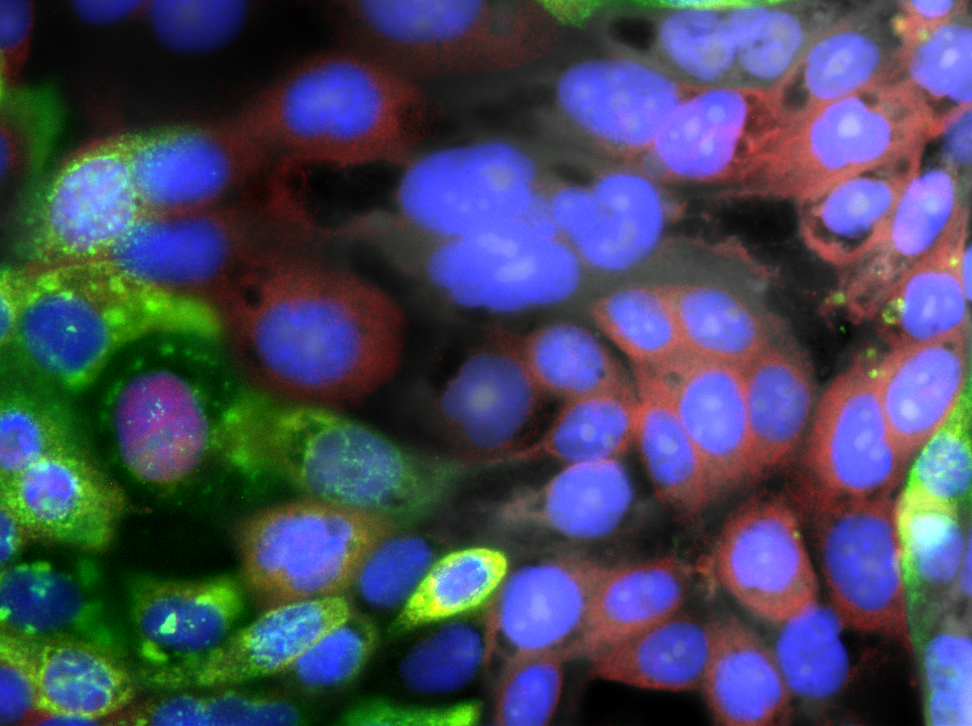
Viral infection means interactions between the virus, the epithelium cells that become infected and the cells involved in the immune response. High content screening (HCS) microscopy is a way of spatially visualizing the infection process when a virus enters a cell, or what happens in the immune response processes that follow, across a large sample size. It thusly becomes possible to see how a virus or other pathogen may bypass or delay the immune response to infect target cells, or how antiviral treatments may affect infection at different stages.
HCS microscopy is a method commonly used in drug discovery to screen for compounds that alter the phenotypes of cells in culture. In virology, measurement of RNA or protein expression can quantify the level of infection. Concomitant imaging of innate immune response processes or cell death markers can be identified and measured to follow the cellular response to infection.
By fluorescently staining cellular structures and targeting fluorescent markers to specific epitopes within the virus/pathogen, HCS can identify cell populations, viral replication and identify intermediates that the virus produces during the infection process. For instance, a recent study of SARS-CoV-2 utilizing HCS identified that upon infection, the virus antagonizes the innate immune response within cells, such that viral replication occurs faster than these cellular protection processes. In this fashion, the virus propagates its infection before the body’s immune system (i.e., macrophages) are activated to destroy infected cells.

Multi-well Screening for Large Sample Sizes
Having large sample sizes, meaning imaging many individual cells, is vital to ensure statistical significance of measured biological changes. Thus, part of the success of HCS in virology to spatially resolve the infection process is the ability to image multi-well plates. Each plate can be prepared with different labels, virus concentrations, antiviral compounds or with varying infection time points (i.e., varying infection exposure times). The ability to then perform imaging over an entire plate permits rapid acquisition of large, informative data sets regarding the infection process.
Virology imaging on large multi-well plates means the instrumentation needs a high degree of automation to reliably capture high-quality images. It must also facilitate extraction of non-biased metrics to permit quantification of trends and tendencies. Cellular process such as translocation (as in the figure 2e below) would be challenging and time-consuming to study without this automation.

Looking to the Future: Super-Resolution in Virology
The nature of light gives rise to the smallest detectable distance between two objects in a microscope, which is commonly in the range of 250-350 nm. Optical physicists refer to this size as the diffraction limit. This means that biological structure or interactions below this length scale are not resolvable. It is particularly relevant for virology, since most viruses are much smaller, being around 100 nm in diameter.
Recent advances in microscopy, however, have enabled creation of techniques capable of resolving biological structure below the diffraction limit. Such methods are termed super-resolution microscopy for their ability to separate structures with length scales ranging from 20-150 nm, depending on the technique, and are appealing for virology.1
Super-resolution microscopy may play a role in the future of virology and the study of viruses, as it is capable of visualizing virus morphology2 and may be helpful in enhancing co-localization of viruses to intracellular compartment relevant for viral replication3.
A challenge for the use of super-resolution techniques is throughput to achieve high-numbers and study large sample sizes. We at IDEA Bio-Medical have innovative solutions that can increase the automation and throughput of some super-resolution methods, as described in the next section.
Automated super-resolution imaging with Hermes system
IDEA Bio-Medical is a joint venture between the Weizmann Institute of Science and IDEA Machine Development, Design and Production. The HERMES platform is a highly flexible high content screening platform. The HERMES is capable of live cell imaging in virology imaging studies and many other tools such as cell counting and cytometry.
Hermes automated imaging enables direct visualization of the intracellular immune response, while preserving the in-situ cellular environment, which is vital to investigating the interplay between the antiviral and inflammatory roles of the innate immune response to viral infection.
Moreover, The HERMES is capable of automated super-resolution imaging using SRRF (super resolution radial fluctuation) to achieve a spatial resolution of 100 nm. The highly automated platform uses algorithms to optimise the focusing and spatial localization to identify cells appropriate for super-resolved imaging within a sample.
To achieve high throughput in the scanning, the HERMES uses a unique design where the objective, rather than the sample stage, is mobile over all three axes. This design features the HERMES with rapid scanning times and ability to scan a 96-well plate in about 100 seconds.
Contact IDEA Bio-Medical today to find out how the HERMES and the ATHENA imaging analysis software could help you with your virology imaging needs and unleash the full power of HCS microscopy.
References
- Arista-Romero, Maria, Silvia Pujals, and Lorenzo Albertazzi. 2021. “Towards a Quantitative Single Particle Characterization by Super Resolution Microscopy: From Virus Structures to Antivirals Design.” Frontiers in Bioengineering and Biotechnology 9(March):1–17. doi: 10.3389/fbioe.2021.647874.
- Arista-Romero M, Pujals S and Albertazzi L (2021) Bioeng. Biotechnol.9:647874. doi: 10.3389/fbioe.2021.647874
- Wang, J, Han, M, Roy, et al. (2022) Cell Reports Methods 2, 100170
DOI 1016/j.crmeth.2022.100170

From Fish to Cure: Discovering Therapies for KLA with Hermes & Athena
April 24, 2025
No Comments
Zebrafish Model Identifies New Drugs for Kaposiform Lymphangiomatosis (KLA) Researchers from the Yaniv Lab at the Weizmann Institute of Science successfully leveraged the Hermes high-content

White Paper: Advancing Therapeutics for Kaposiform Lymphangiomatosis (KLA) with High-Throughput Zebrafish Models
April 24, 2025
No Comments
Explore groundbreaking Kaposiform Lymphangiomatosis (KLA) research using zebrafish models. The Yaniv Lab leverages IDEA Bio-Medical’s Hermes microscope and Athena software for high-throughput drug screening, identifying promising treatments like Cabozantinib and GSK690693 for this rare lymphatic disorder.

Meet us at the Zebrafish Disease Models Society conference in Lisbon!
September 5, 2024
No Comments
The 17th Zebrafish Disease Models Society (ZDMS) annual conference will take place in beautiful Lisbon, Portugal from October 8-10, 2024! Mark your calendars! ZDM meetings
