


From Fish to Cure: Discovering Therapies for KLA with Hermes & Athena
Zebrafish Model Identifies New Drugs for Kaposiform Lymphangiomatosis (KLA) Researchers from the Yaniv Lab at the Weizmann Institute of Science successfully leveraged the Hermes high-content

White Paper: Advancing Therapeutics for Kaposiform Lymphangiomatosis (KLA) with High-Throughput Zebrafish Models
Explore groundbreaking Kaposiform Lymphangiomatosis (KLA) research using zebrafish models. The Yaniv Lab leverages IDEA Bio-Medical’s Hermes microscope and Athena software for high-throughput drug screening, identifying promising treatments like Cabozantinib and GSK690693 for this rare lymphatic disorder.
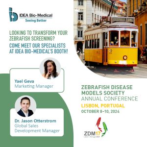
Meet us at the Zebrafish Disease Models Society conference in Lisbon!
The 17th Zebrafish Disease Models Society (ZDMS) annual conference will take place in beautiful Lisbon, Portugal from October 8-10, 2024! Mark your calendars! ZDM meetings