How Spheroid Imaging Factors into Cancer Studies
Creating realistic biological models is a crucial part of medical research for understanding diseases and developing treatments. The use of cell lines to evaluate drug toxicity and treatment potency is standard practice in many areas of medical research, including the development of new cancer therapies.

To date, most research makes use of 2D cell culture lines. A normal 2D cancer cell line will have the tumor cells deposited across a plastic or glass surface like a microscopy slide. While often easy to prepare and use for imaging experiments, the use of these hard flat surfaces as supports for the cells results in surface interactions that differ from the actual 3D biological system and the absence of many critical cell-cell interactions.[2] With developments in cell culturing and bioimaging methods, it is now possible to create 3D cell lines that better imitate the actual in vivo cellular environment. These 3D models are known as spheroids.[1]
While spheroids offer more realistic models and mimics of cancer tumor microenvironments, sometimes performing spheroid imaging can be challenging, with many sample preparation methods for spheroid microscopy proving destructive.[3] Successful spheroid imaging methods approaches need to find ways to suppress scatter, avoid photobleaching and be reasonably quick, for which techniques such as expansion microscopy have worked very well.[3]

Why Spheroid Imaging?
Cancer drug development has a very high failure rate, and using spheroids as more realistic models ensures positive results at the in vivo testing stage translate into successful clinical applications. Many of the initial challenges with high throughput screening of spheroid models for cancer have been overcome. Spheroid imaging has become a viable technique, and a wider range of accurate tumor models have been developed to exploit the additional information in multicellular tumor experiments.4
At IDEA Bio-Medical, we have developed new imaging tools which can help you overcome some of the complexities in spheroid imaging and ensure it can be used as part of routine workflows in target screening.
As well as our range of microscopy imaging platforms, such as the HERMES, we offer unique features to support spheroid imaging. This includes a proprietary laser-based focus dedicated for U-shaped bottom plates, designed explicitly for spheroid imaging which helps with the growth process.
Cellular co-culture in spheroids
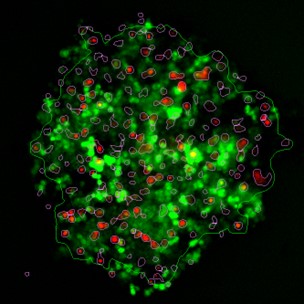
Spheroids are generated by co-culturing tumorigenic cells (green) together with normal, tissue-specific fibroblast cells (red) at different cell seeding densities. Normal cells are visible in the interior/core of the spheroid and tumorigenic cells surrounding them on the outside. The experiment studied tumorigenic cell infiltration into the normal cells as a proxy for metastasis. The screen included compounds reducing the infiltration potential of the tumorigenic cells. Spheroids are identified in the two fluorescence colors and their areas in the image are compared to quantify infiltration of the tumorigenic cells into the normal cells. In this image, the spheroid of normal cells is demarcated with the thick red line and that of the tumorigenic cells with the thin yellow line.


Simplifying Spheroid Imaging
IDEA Bio-Medical offers a wide range of magnifications and fluorescence wavelengths, alongside Z-stack, de-convolution and re-construction image processing tools, to enable flexibility and versatility to support a broad range of experimental designs using spheroid models. Furthermore, our sophisticated imaging and analysis systems incorporate an automated ability to track and record rare events occuring in singular cells within large cell populations, which is a useful feature of great interest in cancer studies. . Another feature of our imaging platforms is imaging and analysis protocols to make an analysis of spheroid imaging data more straightforward, including viability assay recognizing whether the spheroids are live or dead and how the spheroid growth has evolved over the whole plate.

Using the ATHENA software to analyze the results of spheroid imaging experiments allows for automated identification of many of the properties above and high content screening for rapid identification of potential therapeutic targets. The software can be used to quantify and compare different morphological features or signal intensities within each spheroid and related to other spheroids across the plate. Contact us today to find out how our imaging platforms and analysis tools could help you achieve the full power of spheroid imaging. Improve therapeutic development pipelines and cancer research applications with us.
References and further reading:
- Kapałczyńska, M., Kolenda, T., Przybyła, W., Zajączkowska, M., Teresiak, A., Filas, V., Ibbs, M., Bliźniak, R., Łuczewski, Ł., & Lamperska, K. (2018). State of the art paper 2D and 3D cell cultures – a comparison of different types of cancer cell cultures. Arch Med Sci, 4, 910–919. https://doi.org/10.5114/aoms.2016.63743
- Jensen, C., & Teng, Y. (2020). Is It Time to Start Transitioning From 2D to 3D Cell Culture ? Frontiers in Molecular Biosciences, 7, 33. https://doi.org/10.3389/fmolb.2020.00033
- Edwards, S. J., Carannante, V., Kuhnigk, K., Ring, H., Tararuk, T., Hallböök, F., Blom, H., Önfelt, B., & Brismar, H. (2020). High-Resolution Imaging of Tumor Spheroids and Organoids Enabled by Expansion Microscopy. Frontiers in Molecular Biosciences, 7, 208. https://doi.org/10.3389/fmolb.2020.00208
- Labarbera, D. V, Reid, B. G., Yoo, B. H., Labarbera, D. V, Reid, B. G., Hoon, B., & The, Y. (2012). The multicellular tumor spheroid model for high- throughput cancer drug discovery The multicellular tumor spheroid model for high-throughput cancer drug discovery. Expert Opinion on Drug Discovery, 7(9), 819–830. https://doi.org/10.1517/17460441.2012.708334

From Fish to Cure: Discovering Therapies for KLA with Hermes & Athena
Zebrafish Model Identifies New Drugs for Kaposiform Lymphangiomatosis (KLA) Researchers from the Yaniv Lab at the Weizmann Institute of Science successfully leveraged the Hermes high-content

White Paper: Advancing Therapeutics for Kaposiform Lymphangiomatosis (KLA) with High-Throughput Zebrafish Models
Explore groundbreaking Kaposiform Lymphangiomatosis (KLA) research using zebrafish models. The Yaniv Lab leverages IDEA Bio-Medical’s Hermes microscope and Athena software for high-throughput drug screening, identifying promising treatments like Cabozantinib and GSK690693 for this rare lymphatic disorder.

Meet us at the Zebrafish Disease Models Society conference in Lisbon!
The 17th Zebrafish Disease Models Society (ZDMS) annual conference will take place in beautiful Lisbon, Portugal from October 8-10, 2024! Mark your calendars! ZDM meetings
Lorem ipsum dolor sit amet, consectetur adipiscing elit. Ut elit tellus, luctus nec ullamcorper mattis, pulvinar dapibus leo.
