How to Efficiently Conduct 3D Spheroid Imaging
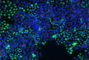
Spheroids are crucial to understanding certain diseases and their cell involvement. These three-dimensional cell aggregates mimic a cellular microenvironment, such as microtumours, and help promote cell-to-cell interactions. Analyzing and imaging spheroids is conducted through 3D spheroid imaging, and the images produced are used to study a range of cellular behaviors. 3D spheroid imaging is a powerful tool used throughout biomedical research to monitor cell interactions, and this blog post will look at how it can be conducted effectively.

Equipment for 3D Spheroid Imaging
3D spheroid imaging requires several pieces of equipment, including consumables such as cell culture flasks and imaging-quality multiwell plates, alongside reagents such as growth media, serum and fluorescence dyes.
On top of that, imaging of 3D models for drug screening, such as Spheroids and Organoids, requires exceptional automated microscopy systems and image analysis capabilities.
High-resolution microscopy is essential for obtaining 3D images. The microscope must support Z-stack acquisition,ability to scan U-shaped bottom plates and post processing tools such as de-convolution and re-construction.
In addition, High resolution to super resolution imaging capabilities in high throughput screening is a unique combination that allows real-time analysis of full biological scales, from whole organelles, through tissues and cells, and down to sub-nuclear nucleic acids.
Many labs may also benefit from flexibility in image load, wherein the same automated microscope supports both single microscopy slide inspections as well as screens of thousands of microplates over long-time experiments with full environmental control for live experiments. Such flexibility allows versatile uses of the same platform, from routine lab inspections such as transfection assays or confluency assays, up to screens of thousands of drug compounds.
Following imaging of the spheroid samples, the images go through automated analysis to extract meaningful, quantitative data from the samples.Various options are available to support Image processing and analysis. This includes software for segmentation, 3D reconstruction, and visualization.
The type of research being conducted dictates the specific equipment needed. Therefore, to efficiently perform spheroid imaging, the scientist will need to choose the most suitable equipment based on the desired outcome. The desired results’ key aspects include accuracy, imagining depth, and image resolution.
IDEA Bio-Medical and Spheroid Imaging
At present, most research uses 2D cell culture lines. Although much can be learned from these 2D images, the option to create 3D cell lines provides significant benefits to the research of diseases and understanding more about cell health and interactions. These 3D cell cultures are the spheroids we discussed in this blog post, which are vital in drug discovery and research into cell culture systems.
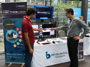
IDEA Bio-Medical has developed a range of spheroid imaging solutions to improve the accuracy and efficiency of spheroid imaging. We offer our HERMES range of microscopes and a selection of features designed to integrate your workflow and enhance imaging applications.
WiSoft Athena
The WiSoft Athena is a ready-made image analysis software designed to easily and quickly evaluate live cell cultures in advanced cell biology applications.
WiScan Hermes
Our WiScan Hermes is a high-content imaging system developed for assay development, compound screens, and analyzing a wide range of cells, such as spheroids. It is an easy-to-use automated system that offers high acquisition speeds and quality images.
Contact IDEA Bio-Medical today for more information on what tools and equipment can be used to conduct 3D spheroid imaging effectively.
References
- Greger, K., Krzic, U., Reynaud, E., and Stelzer, E., (2008) Light sheet-based fluorescence microscopy: more dimensions, more photons, and less photodamage. HFSP J. https://www.ncbi.nlm.nih.gov/pmc/articles/PMC2639947/

What’s next for Zebrafish microscopy in 2023
News: What’s next for Zebrafish microscopy in 2023? Dr. Jason Otterstrom talks about the benefits and challenges of using Zebrafish as an emerging model for

Athena Zfish software- Installer
Time to Elevate Your Research with Athena Zebrafish Software! Dashboard | Technical overview | Pricing Our new, first-of-its-kind analysis software for automated analysis of Zebrafish microscopy images offers simple

Zebrafish: A Preclinical Model for Drug Screening
Zebrafish: A Preclinical Model for Drug Screening Download Software Preclinical models play a fundamental role in the drug discovery pipeline, enabling researchers to identify compounds

What is High Content Imaging?
What is High Content Imaging? High content imaging (HCI), is a powerful image-based paradigm used across the full spectrum of biochemistry, cellular biology, microbiology, molecular
