Biological Applications

Wound Healing / Scratch Assay

Detection of Intracellular Granules / Foci

Quantitive Analysis of Multicolor Labelled Cells

Segmentation of Yeast Cells & Quantifying Their Properties

Colony Formation Assay

3D Object Imaging and Analysis

Wound Healing / Scratch Assay
Tumor Spheroids
Immune Cell Killing
Phagocytosis

Mitochondria Count, Morphology, Protein Expression & More

Counts Fluorescently Labelled Cells or Smaller Objects

Counts Fluorescently Labelled Cells or Smaller Objects

Counting Live & Dead Cell Populations

Quantification of Foci Labelled Intranuclear Proteins

Cytoplasm / Nucleus Protein Expression Ration Quantification

Fluorescence Intensity Quantification

Quantify Cell Population Fraction at Different Cell Cycle Phases

Imaging of the Nematode Caenorhabditis Elegans

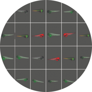
Automated, AI-based analysis of Zebrafish microscopy images

Detect Fibber Structures & Quantify their Length & Intensity

Study changes in cell growth and death, along with spread of the viral infection over time.

Masure Multiple Shape Properties of Fluorescently Labelled Cells or Objects

Measure Confluence Percentage

Fast Automatic Identification and Acquisition of Cell / Object of Interest

Scratch Assay

Wound Healing / Scratch Assay

Intracellular Granules Detection

Detection of Intracellular Granules / Foci

Imaging Cytometry

Quantitive Analysis of Multicolor Labelled Cells

Yeast Colocalization

Segmentation of Yeast Cells & Quantifying Their Properties

Clonogenic Assay

Colony Formation Assay

Confluency Assay

Measure Confluence Percentage

Rare Events Detection

Fast Automatic Identification and Acquisition of Cell / Object of Interest

Spheroids & 3D Imaging

3D Object Imaging and Analysis

Time Lapse Assays

Wound Healing / Scratch Assay
Tumor Spheroids
Immune Cell Killing
Phagocytosis

Mitochondria Analysis​

Mitochondria Count, Morphology, Protein Expression & More

Cell Count in Colonies​

Counts Fluorescently Labelled Cells or Smaller Objects

Cell Count

Counts Fluorescently Labelled Cells or Smaller Objects

Cell Viability-Toxicology Assay​

Counting Live & Dead Cell Populations

Intranuclear Foci​

Quantification of Foci Labelled Intranuclear Proteins

Nuclear Translocation

Cytoplasm / Nucleus Protein Expression Ration Quantification

Protein Expression​

Fluorescence Intensity Quantification

Cell Morphology​

Masure Multiple Shape Properties of Fluorescently Labelled Cells or Objects

Cell Cycle

Quantify Cell Population Fraction at Different Cell Cycle Phases

C-Elegans

Imaging of the Nematode Caenorhabditis Elegans

Zebrafish in-vivo Assays​

High Throughput Imaging Assays using Zebrafish Model

Cytoskeleton Fiber Detection

Detect Fibber Structures & Quantify their Length & Intensity