In the intricate world of biological research, the precision with which one can observe and analyze is paramount. High-resolution microscopy, especially when applied to model organisms like zebrafish, has emerged as an indispensable tool in phenotype identification. This article delves deeper into the nuances of zebrafish microscopy and its profound implications for the broader scientific community.

Zebrafish Microscopy: Unveiling the Microscopic World
Zebrafish, with their transparent embryos and swift developmental stages, have been a cornerstone in biological research for years. Their unique characteristics make them an ideal candidate for microscopic studies. The introduction of high-resolution microscopy, particularly high-content imaging (HCI), has exponentially expanded the horizons of what researchers can discern in zebrafish studies.
HCI’s prowess lies in its ability to provide detailed single-cell measurements, offering a depth of biological specificity and phenotypic complexity previously unattainable. This non-destructive testing approach ensures that the biological integrity of the specimen remains intact, allowing for repeated observations over time.
Furthermore, the integration of computer-driven automation in HCI has been a game-changer. By removing human bias and drastically speeding up repetitive operations, researchers can process and analyze vast datasets in record time. This efficiency is crucial in today’s data-driven research landscape.
Super-Resolution Microscopy: Pushing the Boundaries
While traditional microscopy techniques have served the scientific community well, they come with inherent limitations. Super-resolution microscopy has emerged as a beacon, pushing the boundaries of what’s visually possible. Capable of discerning structures at a resolution of 20-150 nm, this technique is opening doors, especially in virology and infection immunity studies.
The ability to visualize virus morphology in such detail and to enhance the co-localization of viruses to specific intracellular compartments is revolutionizing our understanding of viral replication and behavior. Using Zebrafish models for studying bacteria and viruses, super resolution can shed new light and gain insights never previously explored.
The Future of Zebrafish Research: A Microscopic Revolution
The advancements in microscopy techniques are not merely enhancing our current understanding; they’re setting the stage for future breakthroughs. With the potential applications of these techniques in non-destructive testing and beyond, the future of zebrafish research is poised for a series of revolutionary discoveries.
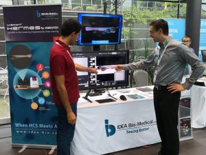
At IDEA Bio, we’ve witnessed the transformative power of high-resolution, automated microscopy in zebrafish research. We firmly believe that these advanced techniques are not just tools but are, in fact, the driving forces of the next wave of scientific innovation.Utilizing high NA oil objectives in full automation on our Hermes high content screening system, enables fully automated super-resolution imaging via SRRF- super resolution radial fluctuations. We invite the global scientific community to join us in this journey. Together, let’s redefine the boundaries of biological research and unlock mysteries previously deemed inscrutable.
References and further reading:
1. Zebrafish skeleton development: High-resolution micro-CT and FIB-SEM block surface serial imaging for phenotype identification. Jeremie Silvent, Anat Akiva,Vlad Brumfeld, Natalie Reznikov, Katya Rechav, Karina Yaniv, Lia Addadi, Steve Weiner.
2. Adam P. Deveau, Victoria L. Bentley, Jason N. Berman. Using zebrafish models of leukemia to streamline drug screening and discovery, Experimental Hematology, Volume 45, 2017, Pages 1-9, ISSN 0301-472X, https://doi.org/10.1016/j.exphem.2016.09.012.

What’s next for Zebrafish microscopy in 2023
News: What’s next for Zebrafish microscopy in 2023? Dr. Jason Otterstrom talks about the benefits and challenges of using Zebrafish as an emerging model for

Athena Zfish software- Installer
Time to Elevate Your Research with Athena Zebrafish Software! Dashboard | Technical overview | Pricing Our new, first-of-its-kind analysis software for automated analysis of Zebrafish microscopy images offers simple

Zebrafish: A Preclinical Model for Drug Screening
Zebrafish: A Preclinical Model for Drug Screening Download Software Preclinical models play a fundamental role in the drug discovery pipeline, enabling researchers to identify compounds

White Paper: Advancing Therapeutics for Kaposiform Lymphangiomatosis (KLA) with High-Throughput Zebrafish Models
Explore groundbreaking Kaposiform Lymphangiomatosis (KLA) research using zebrafish models. The Yaniv Lab leverages IDEA Bio-Medical’s Hermes microscope and Athena software for high-throughput drug screening, identifying promising treatments like Cabozantinib and GSK690693 for this rare lymphatic disorder.
