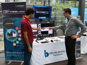
Hermes microscope and Athena image analysis software by IDEA Bio-Medical provide life science researchers with the ability to capture detailed images and perform precise analyses in a rapid and simple fashion. From enhancing tissue regeneration and understanding electromagnetic field impacts to uncovering novel vascular health mechanisms and decoding immune evasion in SARS-CoV-2, these cutting-edge tools are pivotal in advancing high-impact science globally.
Discover how Hermes and Athena are empowering researchers to achieve new heights in scientific discovery and innovation.


One notable study published in the International Journal of Molecular Sciences by a team from Universität Rostock utilized the Hermes imaging system to investigate the effects of Cold Atmospheric Pressure Plasma (CAP) on Mesenchymal Stem/Stromal Cells (MSCs)1. The study demonstrated enhanced MSC migration following CAP treatment, highlighting the therapeutic potential of CAP in tissue regeneration. The Hermes system’s accuracy in wound healing assays was pivotal in monitoring and measuring cellular responses, underscoring its role in advancing regenerative medicine.
Similarly, researchers from UCL- University College London utilized the Hermes and Athena systems in a study published in Nature Microbiology to delve into the immune evasion mechanisms of Omicron subvariants of SARS-CoV-22. The high-resolution fluorescent images captured by the Hermes system and the meticulous quantification of key biomarkers by Athena software were crucial in revealing how these subvariants evade the immune system, providing critical insights for combating the virus.
Further more, researchers from Comenius University in Bratislava employed the Hermes system for advanced fluorescence microscopy in a study published in Nature Scientific Reports. This research explored the effects of electromagnetic fields (EMF) from a novel wireless charging system on human cell lines3. By meticulously analyzing cell morphology, viability, cytotoxicity, genotoxicity, and oxidative stress, the study provided valuable insights into the potential health impacts of EMF exposure amidst the proliferation of wireless technologies.
In another pioneering study by the MIGAL Galilee Research Institute, published in Biochemical and Biophysical Research Communications, the Hermes system, along with Athena software, was used to uncover a novel vascular health mechanism involving an eicosapentaenoic acid-derived lactone oxylipin4. This research demonstrated how the EPA-L metabolite significantly dilates hypertensive arterioles, offering new therapeutic avenues for cardiovascular and metabolic disorders by enhancing endothelial function.
An example for using Hermes and Athena platforms for medical studies using disease models is found in a recent BioRxiv preprint by a group from The Weizmann institute of science. This group presents a novel zebrafish model of Kaposiform Lymphangiomatosis (KLA) that replicates key clinical features of the disease through conditional expression of the NRAS mutation, and demonstrates that Trametinib can reverse these phenotypes5. Using Hermes and Athena AI-based high-throughput drug screening, they identified Cabozantinib and GSK690693 as promising compounds, which were shown to normalize lymphatic endothelial cell (LEC) sprouting and block NRAS pathways in KLA patient-derived cells. This automated screening approach using Zebrafish models significantly accelerate the discovery of potential treatments for KLA.
These examples illustrate the profound impact of Hermes and Athena in high-impact scientific research, enabling significant advancements in understanding complex biological processes and contributing to the development of novel therapeutic strategies worldwide.
Looking to advance into high content screening? Contact us today to see how Hermes & Athena can promote your research.

References:
- Hahn, O.; Waheed, T.O.; Sridharan, K.; Huemerlehner, T.; Staehlke, S.; Thürling, M.; Boeckmann, L.; Meister, M.; Masur, K.; Peters, K. Cold Atmospheric Pressure Plasma-Activated Medium Modulates Cellular Functions of Human Mesenchymal Stem/Stromal Cells In Vitro. J. Mol. Sci.2024, 25, 4944. https://doi.org/10.3390/ijms25094944
- Reuschl, AK., Thorne, L.G., Whelan, M.V.X. et al.Evolution of enhanced innate immune suppression by SARS-CoV-2 Omicron subvariants. Nat Microbiol 9, 451–463 (2024). https://doi.org/10.1038/s41564-023-01588-4.
- Zahumenska, R., Badurova, B., Pavelek, M. et al.Comparison of pulsed and continuous electromagnetic field generated by WPT system on human dermal and neural cells. Sci Rep 14, 5514 (2024). https://doi.org/10.1038/s41598-024-56051-z
- Asulin, M., Gorodetzer, N., Fridman, R., Ben-Shushan, R.S., Cohen, Z., Beyer, A.M., Chuyun, D., Gutterman, D.D. and Szuchman-Sapir, A., 2024. 5, 6-diHETE lactone (EPA-L) mediates hypertensive microvascular dilation by activating the endothelial GPR-PLC-IP3 signaling pathway. Biochemical and Biophysical Research Communications, 700, p.149585. https://doi.org/10.1016/j.bbrc.2024.149585
- Bassi, I., Jabali, A., Farag, N., Egozi, S., Moshe, N., Leichner, G.S., Geva, P., Levin, L., Barzilai, A., Avivi, C. and Long, J., 2024. A high-throughput zebrafish screen identifies novel candidate treatments for Kaposiform Lymphangiomatosis (KLA). bioRxiv, pp.2024-03. https://doi.org/10.1101/2024.03.21.586124

What’s next for Zebrafish microscopy in 2023
News: What’s next for Zebrafish microscopy in 2023? Dr. Jason Otterstrom talks about the benefits and challenges of using Zebrafish as an emerging model for

Athena Zfish software- Installer
Time to Elevate Your Research with Athena Zebrafish Software! Dashboard | Technical overview | Pricing Our new, first-of-its-kind analysis software for automated analysis of Zebrafish microscopy images offers simple

Zebrafish: A Preclinical Model for Drug Screening
Zebrafish: A Preclinical Model for Drug Screening Download Software Preclinical models play a fundamental role in the drug discovery pipeline, enabling researchers to identify compounds

White Paper: Advancing Therapeutics for Kaposiform Lymphangiomatosis (KLA) with High-Throughput Zebrafish Models
Explore groundbreaking Kaposiform Lymphangiomatosis (KLA) research using zebrafish models. The Yaniv Lab leverages IDEA Bio-Medical’s Hermes microscope and Athena software for high-throughput drug screening, identifying promising treatments like Cabozantinib and GSK690693 for this rare lymphatic disorder.
