Using High-Content Imaging in Oncology
High-content imaging, or high-content screening, relies on automated, cutting-edge microscopy technology. Laboratories implementing high-content imaging quickly gain value from their investments in their instruments. They gain time-savings from ease of use for quick user training and fast, autonomous acquisition and analysis to obtain data with large sample sizes.
Oncological research benefits from such automation in assays screening for cancer-related genes, searching for specific cell-types or evaluating cytotoxic properties of a compound library. Such assays can utilize the high-quality images for visualizing and quantifying single cells and intracellular structure, similar to its application in many other areas of biomedical research. This blog post focuses on how researchers may apply high-content imaging in oncology and addresses some solutions provided by leading life sciences companies, such as IDEA Bio-Medical.

Oncological model systems and High Content Imaging
In vitro models are intended to allow researchers to recapitulate aspects of the tumor microenvironment using specific cell types, extracellular matrices, and soluble factors. Traditionally, 2D cell culture of cancer cell lines have been used in high content imaging to identify potential new anti-cancer treatments or identify aberrant phenotypes arising from cancer-related genetic mutations. Such immortalized cell lines, such as the HeLa cells, have provided important insights into oncological cell biology. Recent studies, however, are discovering that the hard, unnatural substrate the cells are grown upon can influence their behavior, giving rise to poor predictions of biological function or compound response in tissues or animal models.
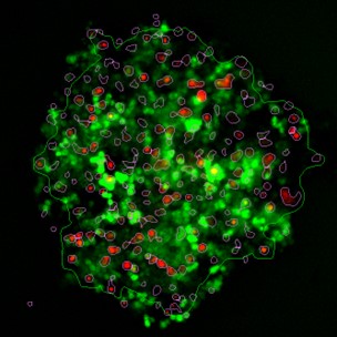
Up to now, moving away from 2D cell culture meant implanting tumor samples into mice. Now, innovative 3D cell culture techniques, such as spheroids and organoids, are bridging the gap between cell in vitro culture and in vivo experiments to study tumor cell behaviour. These methods mimic the 3D physiology within our body to allow for more natural intercellular contacts and inclusion of mechanical effects and chemical signalling into the overarching experimental design.
As well, Zebrafish (Danio rerio) are an emerging animal model for in-vivo cancer studies. The fish are an attractive animal model because of their fast growth with cost-effective maintenance and the possibility of chemical screening in large numbers of animals at reasonable costs. Too, they benefit from amenability to microscopy for easy, direct visualization of implanted human tumor xenografts within the fish for simultaneous evaluation of cell behavior and compound toxicology[1].
High content imaging allows the automated imaging and analysis of all the above mentioned models in one single platform. A wide range of magnifications and fluorescence wavelengths, alongside Z-stack and de-convolution image processing tools, enable flexibility and versatility to support a broad range of experimental designs. Biology can be studied from the sub-cellular range of a few hundreds of nanometres, up to three dimensional structures and whole organisms on the scale of a few millimetres. Furthermore, such sophisticated screening systems have the ability to track and study rare events occurring in only a few cells within a population, a feature particularly suited for studies on diseases like cancer.


The clonogenic assay is a common and sensitive readout for presence of undifferentiated cancer stem cells capable of forming large colonies. Imaging systems can facilitate this assay by digitizing the data as images and analyzing them unbiasedly to measure characteristics like size, density and cell count. The clonogenic assay is an example of how high-content imaging can use bright field images from a label-free sample to automatically identify cells. Such experimental design is also useful for determining cell confluency in a confluency assay without the cost, time, and toxicity associated with traditional staining methods.
Figure 4: Zebrafish models applied for Myelodysplastic syndrome (MDS) and acute myeloid leukemia (AML) studies.
[A]: Image showing bright field and fluorescence channels overlay of a 4dpf zebrafish embryo labeled with CD41:GFP(orange)and lysC:mCherry (cyan) to visualize hematopoietic stem cells and thrombocytes ,and myeloidcells, respectively. Zoom-in highlights the cell types visible in the tail region.[B]: Image from (A) with zebrafish anatomy identified through automatic AI-based image analysis to group the identified cells/ cell clusters found through fluorescence image analysis. Images courtesy of Dr. Alex Lubin, UCL Cancer Institute.

High-content imaging with IDEA Bio
High-content imaging systems by Idea Bio provide users with efficient, user-intuitive, and automated fluorescence microscopy platforms and robust analysis software. The life science solution engineers at IDEA Bio prioritize user needs and develop technologies such as high-content imaging systems, focusing on unique cell biology imaging goals. From specific quantification of intracellular structures to the performance of biochemical assays, high-content imaging systems enable innovative and creative science to address pressing oncological research questions. In particular, IDEA Bio’s WiScan Hermes high-content imaging system increases efficiency and quality of data acquired from laboratory experiments.
If you are interested in using the WiScan Hermes system to address your individualized imaging needs or perhaps need a customized high-content imaging system for specific laboratory uses, please request a demo or reach out to a member of our team today.
References
- Fior, R, Povoa, V, Mndes, RV,et al.PNAS(2017) 114 (39) E8234-E8243. DOI:10.1073/pnas.1618389114

Meet us at the 18th International Zebrafish Conference
June 16, 2024
No Comments
The 18th International Zebrafish Conference (IZFS18) will take place in beautiful Kyoto, Japan from August 17-21, 2024! Mark your calendars! Planning to attend?? Make sure

IDEA Bio-Medical Expands UK Presence with Photon Lines Distribution Partnership
May 29, 2024
No Comments
IDEA Bio-Medical is pleased to announce the appointment of Photon Lines an official distributor in the United Kingdom. As a pioneer in high-content imaging technology,

IDEA Bio-Medical Partners with Bio-Gene Technology to Expand Presence in China
February 6, 2024
No Comments
IDEA Bio-Medical, a leading company specializing in automated microscopy and image analysis systems for life science research, has entered into a strategic distribution agreement with
