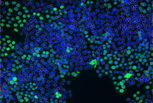
In the realm of biological research, particularly in the study of developmental biology and genetics, the zebrafish (Danio rerio) has emerged as an invaluable model organism. This is primarily due to its genetic similarity to humans, transparent embryos, and rapid development. Image-based assays using zebrafish have revolutionized how researchers observe cellular and molecular processes in-vivo. IDEA Bio-Medical, at the forefront of this technological innovation, utilizes these assays to provide deeper insights into biological phenomena. This article delves into the intricacies of image-based assays in zebrafish research, exploring their applications, methodologies, and the future trajectory of this exciting field.

The Significance of Zebrafish in Biological Research
Zebrafish have become a staple in biological research due to their genetic tractability and embryonic transparency. These features make them particularly suitable for in vivo imaging. With 70% of human genes having at least one zebrafish counterpart, this model organism is instrumental in understanding human biology and disease mechanisms.
Image-Based Assays: A Technological Overview
Image-based assays in zebrafish research involve capturing and analyzing visual data to study biological processes. These assays harness advanced imaging technologies like confocal and/or wide field fluorescence and bright field microscopy, high-throughput imaging, and live imaging techniques. By combining these technologies with fluorescent markers and genetic manipulation tools, researchers can visualize dynamic processes within the whole organism, such as cell proliferation and differentiation, viral infection, and organ development in real-time.
High-Throughput Imaging
High-throughput imaging facilitates the rapid screening of large numbers of zebrafish samples. This approach is vital in drug discovery and toxicological studies, where it is used to assess the effects of various compounds on zebrafish development and physiology.
Live Imaging Techniques
Live imaging techniques enable observing developmental processes as they occur in real-time. This approach provides invaluable insights into the dynamic nature of biological systems.
Applications in Research and Medicine
Image-based assays in zebrafish have broad applications ranging from basic biological research to clinical studies.
Disease Modeling
Zebrafish are increasingly used to model human diseases, including cardiovascular diseases, neurodegenerative disorders, and various forms of cancer. In these models, image-based assays are used to observe disease progression and assess therapeutic compounds’ efficacy in real-time. For example, in cancer research, fluorescently labeled tumor cells can be tracked in live zebrafish, assessing tumor growth, metastasis, and response to anticancer drugs
Drug Discovery
In drug discovery, zebrafish assays offer a cost-effective and ethically viable alternative to mammalian models. They are used to screen potential drug candidates and evaluate their safety and efficacy. This typically occurs via one of two routes:
High-throughput screening (HTS) using zebrafish involves the automated, rapid testing of thousands of chemical compounds to identify potential drugs. In this process, zebrafish are exposed to various compounds, and their responses are monitored using image-based assays. The transparency of zebrafish larvae allows for direct observation of internal processes, such as changes in organ morphology, cellular responses, and behavioral alterations, which can be indicative of a compound’s therapeutic potential or toxicity.
Phenotypic screening in zebrafish allows for the observation of the overall effects of compounds on an organism, rather than targeting a specific molecular pathway. This holistic approach is particularly valuable in identifying compounds with novel mechanisms of action. Image-based assays enable the visualization of phenotypic changes both at the cellular and organism level, providing insights into the compound’s effects on biological systems.
Challenges and Future Directions
While image-based assays using zebrafish provide numerous advantages, they also present challenges, such as sample preparation, data management and the need for advanced image analysis techniques. The future of this field lies in integrating artificial intelligence and machine learning algorithms for enhanced image processing and analysis.
Image-based assays using zebrafish are a powerful tool in biological research, offering unparalleled insights into developmental processes and disease mechanisms. As technology advances, these assays will continue to evolve, further cementing the zebrafish as a model organism of choice for researchers around the globe.
Discover the Future of Zebrafish Research with IDEA-Bio
Ready to delve deeper into the world of zebrafish image analysis? IDEA-Bio is at the forefront of this revolutionary field. Explore our range of products and services designed to enhance your research. Take the first step towards unlocking new possibilities in biological discovery.
References and further reading:
- Lieschke GJ, Currie PD. Animal models of human disease: zebrafish swim into view. Nature Reviews Genetics. 2007;8(5):353-367 1. Lieschke GJ, Currie PD. Animal models of human disease: zebrafish swim into view. Nature Reviews Genetics. 2007;8(5):353-367.
- Kimmel CB, Ballard WW, Kimmel SR, Ullmann B, Schilling TF. Stages of embryonic development of the zebrafish. Developmental Dynamics. 1995;203(3):253-310.
- Sullivan C, Kim CH. Zebrafish as a model for infectious disease and immune function. Fish & Shellfish Immunology. 2008;25(4):341-350.
- Howe K, et al. The zebrafish reference genome sequence and its relationship to the human genome. Nature. 2013;496(7446):498-503.
- Asakawa K, Kawakami K. Targeted gene expression by the Gal4-UAS system in zebrafish. Developmental Growth & Differentiation. 2008;50(6):391-39

What’s next for Zebrafish microscopy in 2023
News: What’s next for Zebrafish microscopy in 2023? Dr. Jason Otterstrom talks about the benefits and challenges of using Zebrafish as an emerging model for

Athena Zfish software- Installer
Time to Elevate Your Research with Athena Zebrafish Software! Dashboard | Technical overview | Pricing Our new, first-of-its-kind analysis software for automated analysis of Zebrafish microscopy images offers simple

Zebrafish: A Preclinical Model for Drug Screening
Zebrafish: A Preclinical Model for Drug Screening Download Software Preclinical models play a fundamental role in the drug discovery pipeline, enabling researchers to identify compounds

What is High Content Imaging?
What is High Content Imaging? High content imaging (HCI), is a powerful image-based paradigm used across the full spectrum of biochemistry, cellular biology, microbiology, molecular
